Introduction
Neural circuits operate through layered codes of ions, metabolites, and peptides that collectively regulate plasticity, stress responses, and circuit homeostasis in experimental preparations. Beyond classical monoamines, neuropeptides signal coordinate gene expression, glial–neuronal cross-talk, energy sensing, and barrier integrity, thereby shaping synaptic wiring and state transitions. These processes intersect with foundational biological problems—oxidative stress, excitotoxicity, protein aggregation, and neuroimmune imbalance—that are frequently modeled in vitro and in preclinical systems to parse causal pathways.
Conventional approaches that globally modulate neurotransmitters can produce diffuse network effects, complicating interpretation in laboratory models. Peptides and peptide-derived fragments, by contrast, often act with circuit and receptor specificity, enabling targeted interrogation of plasticity, cytoprotection, circadian entrainment, and affective state regulation. The sections below reorganize the topic around molecular mechanisms and signaling logic, emphasizing cautious, hypothesis-oriented phrasing and results derived from in vitro studies and preclinical investigations.
Signal Integration and Network Control: Peptides as High-Level Regulators
Peptides participate broadly in neuromodulation, neuroendocrine coupling, and microcircuit gating. Experimental data indicate roles in synaptic transmission, neuronal growth and pruning, blood–brain barrier support, nociceptive processing, and cerebrovascular regulation. Rather than serving as bulk structural elements, they bias how structural features are created, maintained, or removed through matrix enzymes, adhesion complexes, and activity-dependent transcription. Across cultured systems and animal models, 80 endogenous neuropeptides have been cataloged with 100 distinct functional readouts, although many effects arise from peptide–peptide and peptide–monoamine interactions, underscoring the importance of studying combinatorial signaling rather than isolated ligands.
From “Labels” to Mechanisms: A Functional Taxonomy for Experimental Design
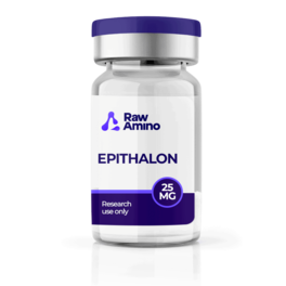
Early discovery often named peptides after their first observed effect, which can obscure the primary CNS roles now apparent in newer models. A mechanism-first taxonomy helps align tools to questions: (i) transcriptional/epigenetic modulators (e.g., short pineal-derived sequences) that alter gene programs; (ii) plasticity amplifiers that interface with neurotrophin pathways; (iii) redox and mitochondrial micropeptides that stabilize bioenergetics; (iv) circadian and immune synchronizers that tune barrier and glial states; and (v) metabolic integrators that couple energy status to memory and motivation. Organizing by intracellular cascades—ERK/CREB, BDNF/TrkB, CaMKII, cAMP/PKA, NF-κB, Nrf2—supports reproducible study designs and interpretable endpoints in vitro and in vivo.
Transcriptional and Synaptogenic Programs: Epithalon, Semax, Selank
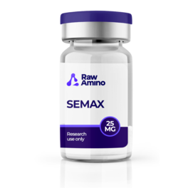
Short sequences such as Epithalon (AEDG) have been reported to influence chromatin accessibility and transcriptional modules linked to neurogenesis and neuronal differentiation in cellular systems, with pineal, retinal, and frontal networks frequently cited as responsive loci. In rodent studies, Semax (an ACTH-derived fragment) increases brain-derived neurotrophic factor (BDNF) in forebrain regions and modulates default-mode and task networks, consistent with facilitation of acquisition/retention through MAPK/ERK and CREB transcriptional pathways. Selank, a tuftsin analog, combines immunomodulatory properties with BDNF-linked plasticity; hippocampal transcriptomics indicate shifts in ion handling and synaptic gene sets. Together, these agents exemplify peptide control over plasticity via convergent regulation of neurotrophins, immediate-early genes, and inhibitory tone, offering tractable readouts (BDNF levels, dendritic spine metrics, activity-regulated transcripts) for laboratory studies.
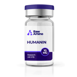
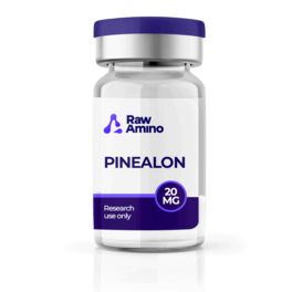
Cytoprotection Under Cellular Stress: Pinealon, Humanin, Noopept, Davunetide
Oxidative stress and excitotoxicity are central constraints in neuronal cultures and rodent models. Pinealon (ultrashort, pineal-derived) has shown antioxidant signatures and protection from excitotoxic injury, with reported upregulation of irisin—a peptide associated with synaptic plasticity—suggesting a myokine–neuron axis relevant to learning paradigms. Humanin, a mitochondria-encoded micropeptide, limits apoptosis by modulating pro-death BCL-2 family signaling and preserving mitochondrial potential; astrocytes release humanin under stress, indicative of glia-to-neuron support that stabilizes synapses in hippocampal systems and retinal epithelium–derived models. Noopept (a dipeptidomimetic) crosses the blood–brain barrier in animal studies, reduces calcium-linked excitotoxic damage, and constrains necrotic zones in ischemia models, enabling quantifiable endpoints such as infarct size and behavioral memory assays in controlled settings. Davunetide (NAP), an eight-mer derived from activity-dependent neuroprotective protein, stabilizes microtubules and has demonstrated neuroprotective and neurotrophic properties in diverse preclinical paradigms; its microtubule-centric mechanism provides clear cytoskeletal readouts (e.g., tau–tubulin interactions, neurite integrity) suitable for imaging-based assays.
Sleep–Endocrine Coupling and Circadian Gateways: DSIP, VIP, Metabolic Peptides
Delta sleep–inducing peptide (DSIP) was initially linked to slow-wave sleep in animal preparations; subsequent work suggests broader coordination of stress axes, nociception, and pituitary rhythms across sleep stages. These effects position DSIP as a candidate for probing how endocrine state gates synaptic consolidation in sleep-dependent memory models. Vasoactive intestinal polypeptide (VIP), while named peripherally, is a central circadian synchronizer via VPAC-receptor signaling, aligning clock gene expression and exerting neurotrophic and anti-inflammatory influences that help preserve endothelial and barrier features in preclinical assays. Metabolic peptides in the ghrelin axis couple energy state to hippocampal plasticity and mesolimbic reinforcement learning, integrating AMPK and ERK pathways with synaptic tagging—useful for experimental paradigms that manipulate energy availability to study memory allocation and reward encoding.
Affective State and Neuropeptides: Vasopressin, Neuropeptide Y, Ghrelin, MIF-1
Affective regulation in laboratory models reflects coordinated changes in monoamines and neuropeptides. Vasopressin and neuropeptide Y (NPY) modulate serotonin/dopamine transmission and stress responsivity, and their expression can shift in response to external neuromodulatory interventions. The ghrelin system influences tryptophan metabolism and links metabolic status to mood-relevant circuitry, motivating experiments that co-vary nutrient context with affective behavior. Melanocyte-inhibiting factor-1 (MIF-1; prolyl-leucyl-glycinamide) illustrates how small peptides may engage dopaminergic receptor systems and intersect with melanocortin signaling; such intersections provide mechanistic entry points to dissect receptor allostery and peptide–monoamine cross-talk using electrophysiology and receptor-binding assays.
Matrix Logic, Barrier Dynamics, and Glial Set-Points
Many peptide effects converge on the extracellular matrix (ECM) and neurovascular unit. By tuning matrix metalloproteinases and their inhibitors, peptides can enable activity-dependent structural remodeling while avoiding maladaptive scarring in brain-adjacent tissues. In parallel, peptide signaling modulates endothelial activation, tight-junction maintenance, and microglial inflammatory tone. Measuring ECM components, barrier permeability markers, and microglial transcriptional states offers multiparametric windows into how peptide programs stabilize or reshape neural microenvironments in controlled experiments.
Conclusion
Across in vitro and preclinical investigations, brain-active peptides function as compact, multifunctional regulators of plasticity, cytoskeletal stability, mitochondrial robustness, circadian entrainment, and neuroimmune balance. Legacy names often understate these roles; mechanism-centric frameworks—anchored in receptor pharmacology, intracellular signaling, and gene-regulatory outcomes—provide clearer hypotheses and reproducible endpoints. Continued laboratory studies that integrate multi-omic profiling, circuit-specific manipulations, and standardized behavioral readouts are warranted to refine causal models of peptide action within neural systems.
References
- T. A. Gudasheva, R. U. Ostrovskaya, and S. B. Seredenin, “Novel Technologies for Dipeptide Drugs Design and their Implantation,” Curr. Pharm. Des., 24(26), 3020–3027, 2018.
- R. H. Ehrensing and A. J. Kastin, “Dose-related biphasic effect of prolyl-leucyl-glycinamide (MIF-I) in depression,” Am. J. Psychiatry, 135(5), 562–566, 1978.
- “A peptide to protect brain function,” ScienceDaily, 2013.
- T. P. Semenova, I. I. Kozlovskiĭ, N. M. Zakharova, and M. M. Kozlovskaia, “[Experimental optimization of learning and memory processes by selank],” Eksp. Klin. Farmakol., 73(8), 2010.
- M. V. Masule et al., “Ghrelin mediated regulation of neurosynaptic transmitters in depressive disorders,” Curr. Res. Pharmacol. Drug Discov., 3, 100113, 2022.
- R. U. Ostrovskaia, “[Evolution of the neuroprotection concept],” Eksp. Klin. Farmakol., 66(2), 32–37, 2003.
- B. H. Morimoto, A. W. Fox, A. J. Stewart, and M. Gold, “Davunetide: a review of safety and efficacy data with a focus on neurodegenerative diseases,” Expert Rev. Clin. Pharmacol., 6(5), 483–502, 2013.
- “Introduction: The Human Brain,” New Scientist, accessed Jan. 10, 2023.
- V. Khavinson et al., “AEDG Peptide (Epitalon) Stimulates Gene Expression and Protein Synthesis during Neurogenesis: Possible Epigenetic Mechanism,” Molecules, 25(3), E609, 2020.
- E. Tuchaai et al., “Deletion of ghrelin alters tryptophan metabolism and exacerbates experimental ulcerative colitis in aged mice,” Exp. Biol. Med., 2022.
- M. B. Segal and B. V. Zlokovic, “The role of peptides in the brain,” in The Blood-Brain Barrier, Amino Acids and Peptides, Springer, 1990, 47–90.
- R. H. Ehrensing, A. J. Kastin, G. F. Wurzlow, G. F. Michell, and A. H. Mebane, “Improvement in major depression after low subcutaneous doses of MIF-1,” J. Affect. Disord., 31(4), 227–233, 1994.
- B. S. Shepherd et al., “Endocrine and orexigenic actions of growth hormone secretagogues in rainbow trout (Oncorhynchus mykiss),” Comp. Biochem. Physiol. A, 146(3), 2007.
- T. A. Kolomin et al., “[Transcriptome alteration in hippocampus under the treatment of tuftsin analog Selank],” Zh. Vyssh. Nerv. Deiat., 63(3), 365–374, 2013.
Disclaimer: The information provided is intended solely for educational and scientific discussion. The compounds described are strictly intended for laboratory research and in-vitro studies only. They are not approved for human or animal consumption, medical use, or diagnostic purposes. Handling is prohibited unless performed by licensed researchers and qualified professionals in controlled laboratory environments.