What Is Humanin?
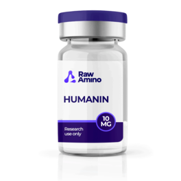
Humanin peptide is one of the more structurally distinctive compounds currently being explored in neuroprotective peptide and mitochondrial peptide research circles. It is a naturally occurring micro-peptide proposed to be encoded in mitochondrial DNA, making it unique among peptides studied in laboratory settings and distinguishing it clearly from the nuclear DNA-encoded compounds that make up the majority of research peptides. This mitochondrial origin has positioned Humanin as a particularly compelling subject of laboratory investigation across cellular protection, neural biology, cardiac tissue, and beyond.
Researchers have suggested that Humanin may exert its biological functions as a cytoprotective protein in laboratory models, with proposed potential to shield cells from apoptosis through the disruption of normal functionality of the Bcl-2-associated X protein (Bax). Research by Yen et al. noted the emerging role of this mitochondrial peptide research subject in stress resistance in laboratory settings, with researchers proposing diverse and intricate mechanisms through which Humanin may demonstrate protective potential across multiple cell types. Different cell types appear to employ distinct receptors to respond to Humanin in laboratory models, with phagocytic cells appearing to express FPRL1 and FPRL2 receptors while neurons may utilize other receptors in these experimental settings.
Humanin Peptide and Neuroprotection Research
At the core of Humanin neuroprotective peptide research is its proposed ability to shield neurons from apoptosis through multiple mechanisms in laboratory models. Research by Matsuoka and by Caricasole et al. suggested that Humanin may exhibit protective interactions against programmed cell death in laboratory settings, extending beyond standard apoptosis to potentially safeguard neurons in the context of neurodegenerative disorder models, counteracting cell death induced by the accumulation of beta-amyloid plaques in these experimental conditions. Researchers also noted potential protection against excitotoxic neuronal death induced by NMDA pulses in laboratory models.
Research by Zhai et al. suggested that Humanin may protect neurons through two distinct proposed mechanisms in laboratory models, both aimed at preventing the activation of the apoptosis pathway within mitochondria. Humanin appeared to bind to Bcl-2 stimulating proteins Bid and tBid in laboratory settings, potentially impeding their function and blocking the initiation of the apoptosis pathway in these models. Research by Zárate et al. further suggested that astrocytes may release Humanin to potentially protect synapses in hippocampal neurons in laboratory settings, with researchers proposing that similar to many natural regulatory processes, Humanin function may decline with age in laboratory models, potentially contributing to age-related observations in neurodegenerative research contexts.
Cardioprotection Research
Building on its neuroprotective research profile, Humanin has also been extensively studied for its proposed cardioprotective interactions in laboratory models. Research by Bachar et al. suggested that Humanin may be present in the walls of blood vessels in laboratory settings, potentially playing a role in safeguarding these vessels from the detrimental impact of oxidized LDL cholesterol. Specifically, Humanin may intervene in the process of LDL oxidation and potentially inhibit the production of reactive oxygen species in response to oxidative stress in laboratory models, with researchers reporting observations of reduced reactive oxygen species levels and decreased apoptosis in these experimental settings.
Research by Cai et al. further suggested that Humanin levels may decline with age in laboratory models, with emerging research indicating that certain disease states may also impact the levels of this mitochondrial peptide research subject. Researchers proposed that Humanin levels may serve as a promising marker in cardiovascular laboratory research contexts, appearing to decline proportionally with the severity of cardiovascular observations in these settings, making cardioprotection one of the most actively documented areas of this neuroprotective peptide’s laboratory research profile.
Humanin Peptide and Retinal Health Research
Beyond its neural and cardiac research profile, Humanin has also been studied for its proposed interactions with retinal tissue in laboratory models. Research by Sreekumar et al. proposed the probable significance of Humanin in the retinal pigment epithelium, a specialized layer of cells in the retina serving crucial functions including light absorption and blood component filtration in laboratory research contexts. In cell culture experiments, supplementation with Humanin appeared to enhance retinal pigment epithelium functionality and resilience against apoptosis in these laboratory settings, with researchers proposing a possible role for this mitochondrial peptide in reducing oxidative stress within ocular tissue in these experimental models.
IGF-1 Interaction Research
One of the more mechanistically distinctive areas of Humanin peptide research involves its proposed interactions with insulin-like growth factor 1 (IGF-1) in laboratory models. Research by Xiao et al. suggested that these two peptides may influence each other in laboratory settings, with Humanin potentially exerting a down-regulatory effect on circulating IGF-1 levels, while IGF-1 may influence Humanin levels in these models. Researchers proposed that in certain laboratory contexts, these peptides may exhibit synergistic interactions, potentially working in conjunction to inhibit apoptosis, moderate inflammation, and support protection against specific forms of heart disease in laboratory settings. Researchers noted that the precise mechanism underlying this interaction remains to be fully elucidated in laboratory models, while acknowledging that Humanin represents an emerging player in IGF-1 signaling research.
Humanin Peptide and Bone Health Research
Rounding out this neuroprotective peptide’s broad laboratory research profile, Humanin has also been explored for its potential interactions with bone and cartilage biology in laboratory models. Research by Celvin et al. suggested that Humanin may exhibit the ability to prevent chondrocyte death in laboratory settings, with chondrocytes considered responsible for producing the collagen matrix important for bone formation in laboratory research contexts. Researchers proposed this interaction may be achieved without compromising the anti-inflammatory properties of glucocorticoids in laboratory models, potentially supporting the growth of bone and cartilage in these settings.
Research by Kang et al. further suggested that Humanin may suppress the formation of osteoclasts in laboratory models, the cells responsible for bone resorption and remodeling in these research contexts. Researchers proposed that by potentially inhibiting osteoclast formation in laboratory settings, Humanin may help to moderate excessive bone remodeling observations in these experimental models, adding a bone health dimension to this mitochondrial peptide research subject’s already expansive laboratory research profile.
References
- Caricasole A, et al. A novel rat gene encoding a Humanin-like peptide endowed with broad neuroprotective activity. FASEB J. 2002;16(10):1331–3.
- Yen K, et al. The emerging role of the mitochondrial-derived peptide Humanin in stress resistance. J Mol Endocrinol. 2013;50(1):R11–9.
- Matsuoka M. Humanin; a defender against Alzheimer’s disease? Recent Pat CNS Drug Discov. 2009;4(1):37–42.
- Caricasole A, et al. A novel rat gene encoding a Humanin-like peptide endowed with broad neuroprotective activity. FASEB J. 2002;16(10):1331–3.
- Xu X, et al. Neuroprotective effect of humanin on cerebral ischemia/reperfusion injury is mediated by a PI3K/Akt pathway. Brain Res. 2008;1227:12–8.
- White AR, et al. Sublethal concentrations of prion peptide PrP106-126 or the amyloid beta peptide activates expression of proapoptotic markers in primary cortical neurons. Neurobiol Dis. 2001;8(2):299–316.
- Zhai D, et al. Humanin binds and nullifies Bid activity by blocking its activation of Bax and Bak. J Biol Chem. 2005;280(16):15815–24.
- Zárate SC, et al. Humanin, a Mitochondrial-Derived Peptide Released by Astrocytes, Prevents Synapse Loss in Hippocampal Neurons. Front Aging Neurosci. 2019;11:123.
- Bachar AR, et al. Humanin is expressed in human vascular walls and has a cytoprotective effect against oxidized LDL-induced oxidative stress. Cardiovasc Res. 2010;88(2):360–6.
- Cai H, et al. Protective Mechanism of Humanin Against Oxidative Stress in Aging-Related Cardiovascular Diseases. Front Endocrinol. 2021;12:683151.
- Sreekumar P, et al. The Mitochondrial-Derived Peptide Humanin Protects RPE Cells From Oxidative Stress, Senescence, and Mitochondrial Dysfunction. Invest Ophthalmol Vis Sci. 2016;57:1238.
- Xiao J, et al. Humanin: Functional Interfaces with IGF-I. Growth Horm IGF Res. 2016;29:21–27.
- Celvin B, et al. Humanin prevents undesired apoptosis of chondrocytes without interfering with the anti-inflammatory effect of dexamethasone. Clin Exp Rheumatol. 2020;38(1):129–135.
- Kang N, et al. Humanin suppresses receptor activator of nuclear factor-κB ligand-induced osteoclast differentiation via AMPK activation. Korean J Physiol Pharmacol. 2019;23(5):411–417.
Disclaimer: The information provided is intended solely for educational and scientific discussion. The compounds described are strictly intended for laboratory research and in-vitro studies only. They are not approved for human or animal consumption, medical use, or diagnostic purposes. Handling is prohibited unless performed by licensed researchers and qualified professionals in controlled laboratory environments.