
What Is PEG-MGF?
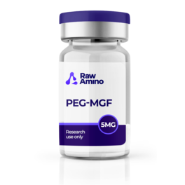
PEG-MGF peptide, formally known as polyethylene glycol modified Mechano Growth Factor, is one of the more structurally distinctive compounds currently being explored across muscle regeneration peptide and tissue repair research circles. It is a synthetic peptide derived from Mechano Growth Factor (MGF), itself a notable variant of insulin-like growth factor 1 (IGF-1) that emerges as a potential mediator involved in tissue growth and repair processes across diverse physiological systems including muscle, tendon, cardiac, cerebral, and skeletal structures.
What sets PEG-MGF apart from standard MGF in laboratory research is the addition of polyethylene glycol (PEG), a biocompatible polymer, to the MGF peptide structure through a process called PEGylation. This modification is speculated by researchers to optimize the peptide’s delivery and bioavailability in laboratory settings, addressing one of the key limitations of standard MGF — its rapid degradation in biological environments. The inclusion of PEG is also proposed to enhance water solubility and stability, potentially decrease the molecule’s clearance through the kidneys, and extend its circulation time in laboratory models. This combination of structural stability and broad tissue research applicability has made PEG-MGF a particularly active subject of laboratory investigation across multiple biological systems.
Why PEGylation Matters: The Science Behind the Modification
To understand what makes this mechano growth factor peptide research subject distinctive, it helps to appreciate what PEGylation achieves in laboratory settings. Standard MGF degrades rapidly in biological environments, limiting its usefulness as a research tool for studying prolonged or systemic tissue interactions. By attaching polyethylene glycol to the MGF molecule, researchers created a compound with meaningfully different pharmacokinetic properties in laboratory models.
PEGylation is a well-established approach in peptide research for several reasons. Beyond extending circulation time, it is thought to reduce immunogenic responses and improve the water solubility of peptides that might otherwise be difficult to work with in laboratory settings. For a compound like MGF, whose expression naturally responds to various physiological stimuli and orchestrates cellular proliferation and migration across multiple tissue types, the ability to study it over extended periods in laboratory models represents a meaningful research advantage. This has opened up investigation across a remarkably broad range of tissue systems, from skeletal muscle and cardiac tissue to bone, cartilage, dental structures, and neural tissue.
PEG-MGF Peptide and Skeletal Muscle Regeneration
At the core of PEG-MGF peptide research is its proposed interactions with skeletal muscle repair in laboratory models. Research by Liu et al. suggested that MGF exposure into injured muscles may confer protection by potentially moderating inflammatory hormone expression and oxidative stress in laboratory settings. Skeletal muscle injuries are among the most prevalent research contexts for this muscle regeneration peptide, with the intricate process of regeneration following injury making it a compelling laboratory model for studying tissue repair mechanisms.
Further investigations into MGF’s potential role in muscle inflammation following skeletal muscle injury suggested a possible regulatory role in laboratory settings, spurring ongoing research into how PEG-MGF’s extended bioavailability might influence these inflammatory dynamics over longer experimental timeframes.
PEG-MGF Peptide and Cardiac Repair Research
Beyond skeletal muscle, PEG-MGF has also been studied for its potential interactions with cardiac tissue repair in laboratory models. Research from the Department of Bioengineering at the University of Illinois suggested that PEG-MGF may exhibit inhibitory interactions with programmed cell death in cardiac muscle subjected to hypoxia in laboratory settings. MGF also appeared to facilitate the recruitment of cardiac stem cells to sites of simulated injury in these models, potentially supporting cardiac regeneration following myocardial infarction in laboratory conditions.
Research by Peña et al. further suggested that localized exposure of PEG-MGF may contribute to enhanced cardiac function following simulated myocardial infarction in laboratory models by moderating pathological hypertrophy and promoting favorable hemodynamic outcomes. Researchers proposed that cardiac-restricted delivery of the MGF E-domain peptide via polymeric microstructures may be relevant to studying adverse remodeling of the heart in laboratory settings.
PEG-MGF Peptide and Bone Repair Research
PEG-MGF has also been studied for its potential interactions with bone repair processes in laboratory models. Research by Deng et al. using rabbit models suggested that PEG-MGF exposure may expedite bone repair by potentially augmenting osteoblast proliferation, the cells responsible for bone mineralization in laboratory settings. The reported healing observations in these rabbit models suggested a possible role for PEG-MGF in bone repair mechanisms, making it an active subject of orthopedic tissue research in controlled laboratory environments.
PEG-MGF Peptide and Cartilage Protection Research
Closely related to its bone research profile, PEG-MGF has also drawn interest for its potential interactions with cartilage biology in laboratory models. Research by Xu et al. suggested that MGF may potentially enhance chondrocyte function in laboratory settings, with studies in murine models proposing that PEG-MGF peptide may facilitate the migration of chondrocytes from bone to cartilage. Researchers further noted that the activity of the p38 MAPK pathway in cells cultured under mechanical overload appeared to be decreased by MGF peptide addition in laboratory models, suggesting that MGF may help moderate mechanical overload-induced cell apoptosis through this signaling pathway in these experimental settings.
PEG-MGF Peptide and Dental Research
One of the more specialized areas of PEG-MGF peptide research involves its potential interactions with periodontal ligament cells in laboratory models. In vitro studies suggested that PEG-MGF may enhance the expression of ligament repair factors including MMP-1 and MMP-2, considered essential for maintaining tooth stability within the bone in laboratory tissue models. Research by Chen et al. proposed that MGF may regulate cyclic stretch-induced osteogenic differentiation and MMP expression through the MEK/ERK1/2 pathway in human periodontal ligament cells in laboratory settings, suggesting a potential avenue for further study in dental tissue research.
PEG-MGF Peptide and Neuroprotection Research
Rounding out this mechano growth factor peptide’s remarkably broad tissue research profile is its proposed potential in neural tissue research. Multiple laboratory investigations utilized murine models to examine elevated MGF concentrations on brain cells, with one study involving mice overexpressing MGF within the hippocampal region, a brain area primarily associated with regulating neurogenesis in laboratory models. These studies reported elevated concentrations of BrdU, a recognized biological marker indicative of proliferative activity, in these laboratory models.
Research by Windebank and colleagues at the Mayo Clinic suggested that increasing MGF levels may potentially promote the growth of new brain cells in regions associated with brain renewal in laboratory models. Higher MGF levels in transgenic laboratory mice appeared to confer resistance to age-related brain damage and improved cognitive function in these models, suggesting a possible neuroprotective role for MGF in laboratory research addressing age-related neuron loss. These findings have added a compelling neural dimension to this muscle regeneration peptide’s already expansive multi-tissue research profile in controlled laboratory environments.
References
- Zabłocka B, et al. Mechano-Growth Factor: an important cog or a loose screw in the repair machinery? Front Endocrinol. 2012.
- Hamley IW. PEG-Peptide Conjugates. American Chemical Society. 2014.
- Janssen JA, et al. Potency of Full-Length MGF to Induce Maximal Activation of the IGF-I R. PLoS One. 2016;11(3):e0150453.
- Liu X, et al. Impaired Skeletal Muscle Regeneration Induced by Macrophage Depletion Could Be Partly Ameliorated by MGF Injection. Front Physiol. 2019;10:601.
- Xu Q, et al. Mechano growth factor attenuates mechanical overload-induced nucleus pulposus cell apoptosis through inhibiting the p38 MAPK pathway. Biosci Rep. 2019;39(3):BSR20182462.
- Doroudian G, et al. Sustained delivery of MGF peptide from microrods attracts stem cells and reduces apoptosis of myocytes. Biomed Microdevices. 2014;16(5):705–15.
- Peña JR, et al. Localized delivery of mechano-growth factor E-domain peptide via polymeric microstructures improves cardiac function following myocardial infarction. Biomaterials. 2015;46:26–34.
- Deng M, et al. Mechano growth factor E peptide promotes osteoblasts proliferation and bone-defect healing in rabbits. Int Orthop. 2011;35(7):1099–106.
- Xu Q, et al. Mechano growth factor attenuates mechanical overload-induced nucleus pulposus cell apoptosis. Biosci Rep. 2019;39(3):BSR20182462.
- Chen JT, et al. Mechano-growth factor regulated cyclic stretch-induced osteogenic differentiation and MMP-1, MMP-2 expression in human periodontal ligament cells. Shanghai Kou Qiang Yi Xue. 2019;28(1):6–12.
- Walker A. Hearts and Minds of Mice and Men: Mechano Growth Factor a new tool in the battle against age-related neuron loss? 2017.
Disclaimer: The information provided is intended solely for educational and scientific discussion. The compounds described are strictly intended for laboratory research and in-vitro studies only. They are not approved for human or animal consumption, medical use, or diagnostic purposes. Handling is prohibited unless performed by licensed researchers and qualified professionals in controlled laboratory environments.