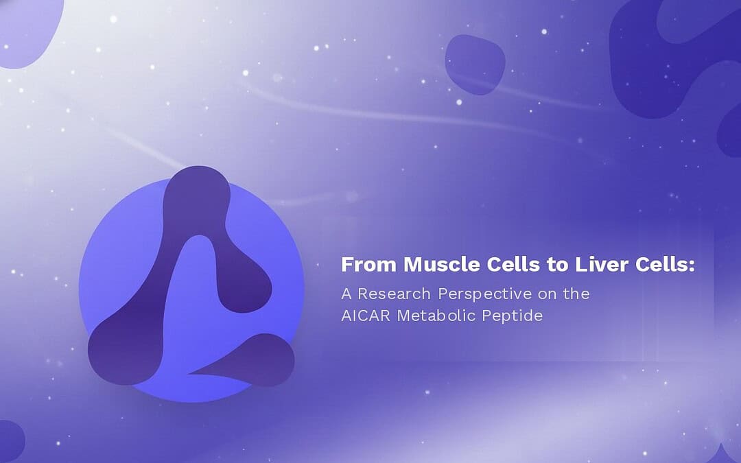
What Is AICAR?
AICAR peptide — formally known as 5-Aminoimidazole-4-carboxamide ribonucleotide — is one of the more broadly studied compounds in cellular energy metabolism research. It is a synthetic analog of adenosine monophosphate (AMP), a naturally occurring nucleotide that plays a key role in storing and transferring energy within cells. Because AICAR mimics AMP, it is thought to activate an enzyme called AMP-activated protein kinase (AMPK) — a molecular sensor widely recognized in research settings for its role in helping cells maintain energy balance while limiting excessive energy storage.
By stimulating AMPK in laboratory models, this energy metabolism peptide has been proposed to exert a wide range of potentially relevant cellular effects — including supporting glucose availability in muscle cells, promoting the breakdown of stored energy under stress conditions, and reducing lipid accumulation in liver cells. AICAR may also influence fatty acid oxidation, mitochondrial biogenesis, and the maintenance of fatigue-resistant muscle fiber characteristics in laboratory models — making it one of the more multifaceted metabolic peptides currently under active laboratory investigation.
AICAR Peptide and Muscle Cell Metabolism
One of the most actively studied areas of AICAR peptide research involves its potential interactions with glucose metabolism in muscle cells — a process that sits at the intersection of cellular energy management and insulin signaling research.
Research by Cuthbertson et al. proposed that AICAR peptide may support muscle cell insulin sensitivity through mechanisms related to its intracellular conversion into a compound called ZMP (5-aminoimidazole-4-carboxamide ribonucleoside). ZMP is thought to resemble AMP in its ability to bind and activate AMPK — and once AICAR enters a muscle cell and is metabolized into ZMP, researchers have hypothesized that a downstream signaling cascade may be initiated that supports the relocation and functionality of glucose transporters. This may potentially allow more efficient glucose uptake in muscle cells under laboratory conditions.
However, it is worth noting that Cuthbertson et al. also referenced studies where AICAR peptide appeared to lead to minimal or no AMPK activation, despite an apparent rise in glucose uptake — suggesting that alternative pathways, including those involving ERK1/2 phosphorylation, may also play a role in mediating glucose transport in muscle cells in laboratory settings. The precise relationship between AICAR, AMPK, and glucose handling therefore remains an area of active investigation.
Research by Boon et al. also observed higher levels of ACC phosphorylation following AICAR peptide exposure in laboratory models — a finding that may reflect activation of a pathway that reduces malonyl-CoA levels and encourages fatty acid oxidation rather than storage in muscle cells. Researchers have proposed that this shift in lipid usage might partly explain how AICAR peptide could influence muscle cell insulin sensitivity — though ongoing experiments continue to evaluate the full scope of this potential mechanism.
AICAR Peptide and Muscle Cell Endurance
Beyond its interactions with glucose metabolism, AICAR peptide has also been explored for its potential influence on muscle cell endurance and performance in laboratory models — an area that has attracted considerable interest in energy metabolism peptide research.
Research by Narkar et al. proposed that by activating AMPK, AICAR peptide may potentially interact with a nuclear receptor called PPARδ — a regulator of genes involved in lipid metabolism and energy expenditure. When activated alongside its co-regulators, PPARδ may coordinate the expression of multiple genes tied to fatty acid oxidation, mitochondrial biogenesis, and the maintenance of slow-twitch, fatigue-resistant muscle fibers in laboratory models.
In laboratory experiments, AICAR peptide appeared to boost the expression of several oxidative metabolism-associated genes — including Ppargc1a, which encodes PGC1α, a co-activator associated with mitochondrial formation and function, and Pdk4, which may regulate glucose utilization in cells. Researchers described this as a metabolic state potentially resembling repeated mechanical stimulation — with the peptide appearing to reprogram muscle cell gene networks in the absence of physical stimulation in laboratory settings. Narkar et al. specifically noted that four weeks of AICAR peptide exposure appeared to induce metabolic gene expression changes and support running endurance by approximately 44% in the laboratory models studied.
Research by Bosselaar further suggested that AICAR peptide may exert a vasodilatory effect on muscle tissue vasculature in laboratory models — possibly through mechanisms involving nitric oxide (NO) — with observations of time- and concentration-dependent increases in blood flow. Researchers proposed that this better-supported blood flow to muscle cells may further contribute to their endurance-related properties in laboratory studies — adding a vascular dimension to this metabolic peptide’s muscle cell research profile.
AICAR Peptide and Cardiac Cell Research
Moving beyond skeletal muscle, AICAR peptide has also been studied for its potential interactions with cardiac cells — particularly in laboratory models of ischemia and cardiac cell stress. This represents one of the more nuanced areas of AICAR energy metabolism peptide research, with researchers exploring both its potential energy-supporting and tissue-organizing properties in compromised cardiac cell environments.
Research by Longnus et al. suggested that AICAR peptide and its active intracellular form ZMP may promote glycogen breakdown in cardiac cells — potentially providing greater ATP availability during ischemic or otherwise stressful conditions in laboratory models. By allosterically activating glycogen phosphorylase, ZMP may support glycogenolysis — a process that could help sustain energy production when oxygen availability is limited. Researchers proposed that this, combined with other AMPK-mediated processes such as the potential relief of malonyl-CoA-induced inhibition on fatty acid transport, may create a dual energy production pathway — drawing on both glucose and fatty acid sources — potentially helping cardiac cells maintain ATP levels in laboratory stress models.
Research by Cieslik et al. further explored AICAR peptide’s potential interactions with fibroblast precursor cells in cardiac tissue models — suggesting that AMPK activation by AICAR may help drive these precursor cells toward becoming functional fibroblasts and, later, myofibroblasts following cardiac cell injury in laboratory settings. Researchers proposed that by supporting fibroblast differentiation and their capacity to produce collagen and α-SMA, AICAR peptide may contribute to forming a more organized extracellular matrix in the healing region — potentially reducing the mechanical stresses that might otherwise worsen damage in compromised cardiac tissue models.
AICAR Peptide and Liver Cell Metabolism
Rounding out this energy metabolism peptide’s broad cellular research profile, AICAR has also been studied for its potential interactions with fat accumulation and lipid metabolism in liver cells in laboratory settings.
Research by Tomita et al. suggested that via AMPK-related mechanisms, AICAR peptide may be able to decrease the activity of specific lipogenic regulators in liver cells — potentially reducing fat accumulation in these models. One proposed mechanism involves AICAR peptide reducing the expression of SREBP-1c — a major transcription factor controlling de novo lipogenesis — which might in theory curb downstream enzymes such as fatty acid synthase and limit the production of new fat molecules in liver cells.
Researchers also observed that AICAR peptide appeared to decrease the products of lipid peroxidation in these liver cell models — with researchers suggesting this may have occurred because a lower triglyceride burden leads to fewer lipid-derived oxidative byproducts. Since lipid peroxidation is often linked to progressive cellular stress in laboratory models, a decrease in these reactive byproducts may potentially lessen subsequent cellular injury. Researchers also noted that additional pathways — including interactions with Kupffer cells, the liver’s resident immune cells — are thought to be involved in AICAR’s interactions with liver cell biology, representing a promising avenue for further laboratory investigation into this metabolic peptide’s hepatic research profile.
References
- Cuthbertson DJ, et al. 5-aminoimidazole-4-carboxamide 1-beta-D-ribofuranoside acutely stimulates skeletal muscle 2-deoxyglucose uptake in healthy men. Diabetes. 2007;56(8):2078–84.
- Boon H, et al. Intravenous AICAR administration reduces hepatic glucose output and inhibits whole-body lipolysis in type 2 diabetic patients. Diabetologia. 2008;51(10):1893–900.
- Narkar VA, et al. AMPK and PPARdelta agonists are exercise mimetics. Cell. 2008;134(3):405–15.
- Bosselaar M, et al. Intra-arterial AICA-riboside administration induces NO-dependent vasodilation in vivo in human skeletal muscle. Am J Physiol Endocrinol Metab. 2009;297(3):E759–66.
- Longnus SL, et al. 5-Aminoimidazole-4-carboxamide 1-beta-D-ribofuranoside stimulates myocardial glycogenolysis by allosteric mechanisms. Am J Physiol Regul Integr Comp Physiol. 2003;284(4):R936–44.
- Cieslik KA, et al. AICAR-dependent AMPK activation improves scar formation in the aged heart in a murine model of reperfused myocardial infarction. J Mol Cell Cardiol. 2013;63:26–36.
- Tomita K, et al. AICAR, an AMPK activator, has protective effects on alcohol-induced fatty liver in rats. Alcohol Clin Exp Res. 2005;29(12 Suppl):240S–5S.
Disclaimer: The information provided is intended solely for educational and scientific discussion. The compounds described are strictly intended for laboratory research and in-vitro studies only. They are not approved for human or animal consumption, medical use, or diagnostic purposes. Handling is prohibited unless performed by licensed researchers and qualified professionals in controlled laboratory environments.